Corneal Mapping
Corneal topography, also known as photokeratoscopy or videokeratography, is a non-invasive medical imaging technique for mapping the surface curvature of the cornea, the outer structure of the eye. Since the cornea is normally responsible for some 70% of the eye's refractive power, its topography is of critical importance in determining the quality of vision.
The three-dimensional map is, therefore, a valuable aid to the examining ophthalmologist or optometrist and can assist in the diagnosis and treatment of a number of conditions; in planning refractive surgery such as LASIK and evaluation of its results; or in assessing the fit of contact lenses. A development of keratoscopy, corneal topography extends the measurement range from the four points a few millimeters apart that is offered by keratometry to a grid of thousands of points covering the entire cornea. The procedure is carried out in seconds and is completely painless.
Anterior Segment Camera KOWA
While there are many digital cameras available, often an anterior segment camera can provide a digital photograph of the eye with the proper color, contrast, sensitivity, and resolution easily by an eye doctor. By getting a clear image of the front of the eye, this information can help diagnose irregularities with the cornea, misalignments, and various eye conditions or damage that may affect your eyes on an external level.
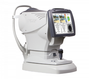 Wavefront Analysis with Corneal Topography- Marco 3D Wavefront
It’s no surprise that no pair of eyes are the same, but aside from color, shape, and health, every eye has a unique surface that determines how clearly you view the world. If you’ve ever wondered why you had a strong prescription, your genetic makeup is a big player, and how your eye is shaped and formed can mean the difference between a new pair of glasses, toric contact lenses, or a pair of scleral lenses for irregular corneas. Our Marco 3D Wavefront captures a complete contour map of your eye’s surface to help our optometrist select the optimal solution for your vision.

Scout Topographer
Similar to the Marco Wavefront, the Scout Topographer is a smaller version that captures your eye’s cornea shape & unique surface structure. The Scout provides Dr. Schwartz another avenue to review your particular visual needs in choosing the exact eyewear solution or specialty contact lens.
Digital Retinal Imaging & OCT Scans
We use cutting-edge digital imaging technology to assess your eyes. Many eye diseases, if detected at an early stage, can be treated successfully without total loss of vision. Your retinal Images will be stored electronically. This gives the eye doctor a permanent record of the condition and state of your retina.
This is very important in assisting your Optometrist to detect and measure any changes to your retina each time you get your eyes examined, as many eye conditions, such as glaucoma, diabetic retinopathy, and macular degeneration are diagnosed by detecting changes over time.
The advantages of digital imaging include:
- Quick, safe, non-invasive and painless
- Provides detailed images of your retina and sub-surface of your eyes
- Provides instant, direct imaging of the form and structure of eye tissue
- Image resolution is extremely high quality
- Uses eye-safe near-infra-red light
- No patient prep required
Digital Retinal Imaging
Digital Retinal Imaging allows your eye doctor to evaluate the health of the back of your eye, the retina. It is critical to confirm the health of the retina, optic nerve, and other retinal structures. The digital camera snaps a high-resolution digital picture of your retina. This picture clearly shows the health of your eyes and is used as a baseline to track any changes in your eyes in future eye examinations.
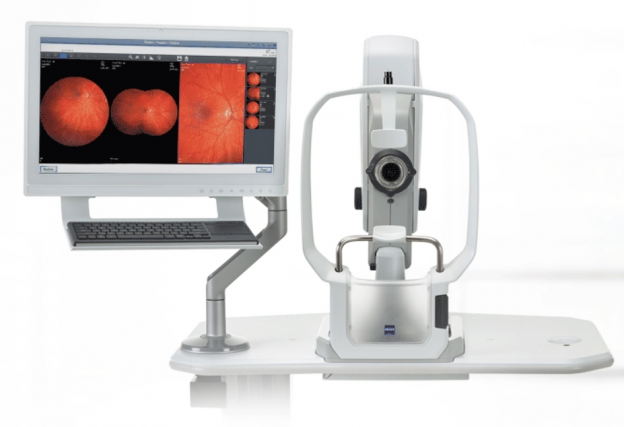 ZEISS CLARUS 500 Fundus Camera
The new leading technology in digital retinal imaging is the Clarus 500 fundus camera by Zeiss. This fundus camera is a new ultra-widefield retinal camera to allow precise color imagery of the eye during a comprehensive eye exam. The color adds additional detail to a clear map of your eye health. Our optometrists, Dr. Moshe Schwartz and Dr. Jenny Chen, include the imaging to diagnose and document any early signs of ocular disease. The CLARUS 500 can produce an image of the macula and periphery of the eye, which is essential in managing macular degeneration. Instead of relying on eye dilations to get a picture of your eye, our new fundus camera creates an amazing blueprint of your eye instantly, so you get the full view of your eye health comfortably and without the drops.
 Eye Check from Volk
Measuring your Volk Eye Check can be used as a routine complementary diagnostic and data gathering tool, where measurements can be gathered and recorded to track your ocular health over time. Great for children or a simple analysis for time-sensitive patients, the Volk Eye Check’s consistent outputs provide objective accurate measurements, aids in the detection of other abnormalities, and even provides an electronic & printable record of your eye’s measurements. The main feature is that these measurements are also useful for contact lens fittings for those who seek specialty contact lenses. Our office makes use of the efficiency of the Volk Eye Check to track the status of your eye shape so that we’ll provide you the best in contact lens care.
Optical Coherence Tomography (OCT)
An Optical Coherence Tomography scan (commonly referred to as an OCT scan) is the latest advancement in imaging technology. Similar to ultrasound, this diagnostic technique employs light rather than sound waves to achieve higher resolution pictures of the structural layers of the back of the eye.
A scanning laser used to analyze the layers of the retina and optic nerve for any signs of eye disease, similar to a CT scan of the eye. It works using light without radiation and is essential for early diagnosis of glaucoma, macular degeneration, and diabetic retinal disease.
With an OCT scan, doctors are provided with color-coded, cross-sectional images of the retina. These detailed images are revolutionizing early detection and treatment of eye conditions such as wet and dry age-related macular degeneration, glaucoma, retinal detachment and diabetic retinopathy.
An OCT scan is a noninvasive, painless test. It is performed in about 10 minutes right in our office. Feel free to contact our office to inquire about an OCT at your next appointment.
Zeiss OCT Cirrus HD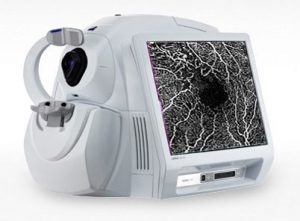
To obtain images of the deepest part of the eye’s structure, an OCT or optical coherence tomography accurately captures a magnified image of the layers of the retina. These images help our eye doctor detect ocular diseases like macular degeneration or glaucoma. These scans are so precise and at such a sophisticated level that an optometrist can identify each specific sign of deterioration and immediately formulate the exact treatment needed to prevent vision loss.
Visual Field Testing
A visual field test measures the range of your peripheral or “side” vision to assess whether you have any blind spots (scotomas), peripheral vision loss or visual field abnormalities. It is a straightforward and painless test that does not involve eye drops but does involve the patient's ability to understand and follow instructions.
An initial visual field screening can be carried out by the optometrist by asking you to keep your gaze fixed on a central object, covering one eye and having you describe what you see at the periphery of your field of view. For a more comprehensive assessment, special equipment might be used to test your visual field. In one such test, you place your chin on a chin rest and look ahead. Lights are flashed on, and you have to press a button whenever you see the light. The lights are bright or dim at different stages of the test. Some of the flashes are purely to check you are concentrating. Each eye is tested separately and the entire test takes 15-45 minutes. These machines can create a computerized map out your visual field to identify if and where you have any deficiencies.
VEP (Visually Evoked Potential Testing)
We can measure your visual pathway through light-evoked signals, which tells how your central vision is functioning. The waveforms of your visual pathway can help Dr. Schwartz diagnose refractive error, vision disorders like amblyopia, and even the health of the optic nerve.
 Marco RT900 and TRS-5100 Digital refractors
Calculation your next glasses prescription ought to be blazing fast & as accurate as possible. While eye doctors have relied on good judgment, Eyesymmetry Vision Center boasts the Marco TRS-5100 & RT900 to provide you the best in digital refraction technology. The small, lightweight unit shouldn’t fool - this piece of hardware will provide your eye doctor with immediate results about your prescription with the utmost accuracy. There’s no better technology in eye care that can determine your exact visual needs than the Marco.

